
Valvular Heart
Disease & TAVR
Valvular Guide Contents
Experiencing Shortness of Breath or Murmur?
Book EvaluationExperiencing Shortness of Breath or Murmur?
What is Valvular Heart Disease?
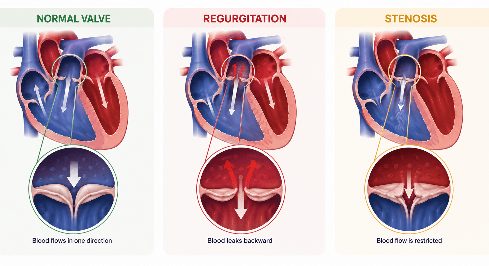
Your heart features four essential valves — Aortic, Mitral, Tricuspid, and Pulmonary — that open and close with every heartbeat to ensure blood flows strictly forward through the heart chambers to the lungs and body.
Valvular heart disease develops when valves become calcified and stiffened (Stenosis), preventing full opening, or when valve leaflets fail to seal tightly (Regurgitation / Insufficiency), allowing blood to leak backward into the heart.
Over time, untreated valve disease can strain the heart and lead to complications like heart enlargement, rhythm problems, or heart failure.
Symptoms of Valve Failure
Dizziness & Fainting (Syncope)
Sudden lightheadedness or fainting caused by temporary reduction of blood flow to the brain across a severe aortic valve stenosis.
Heart Palpitations & AFib
Fluttering or irregular heartbeats resulting from atrium stretching caused by valve regurgitation.
Ankle & Foot Swelling (Edema)
Fluid retention in lower extremities due to right-sided heart congestion and elevated venous pressure.
Chronic Fatigue & Weakness
Overwhelming tiredness caused by reduced cardiac output delivered to body organs.
Diagnostic Tests
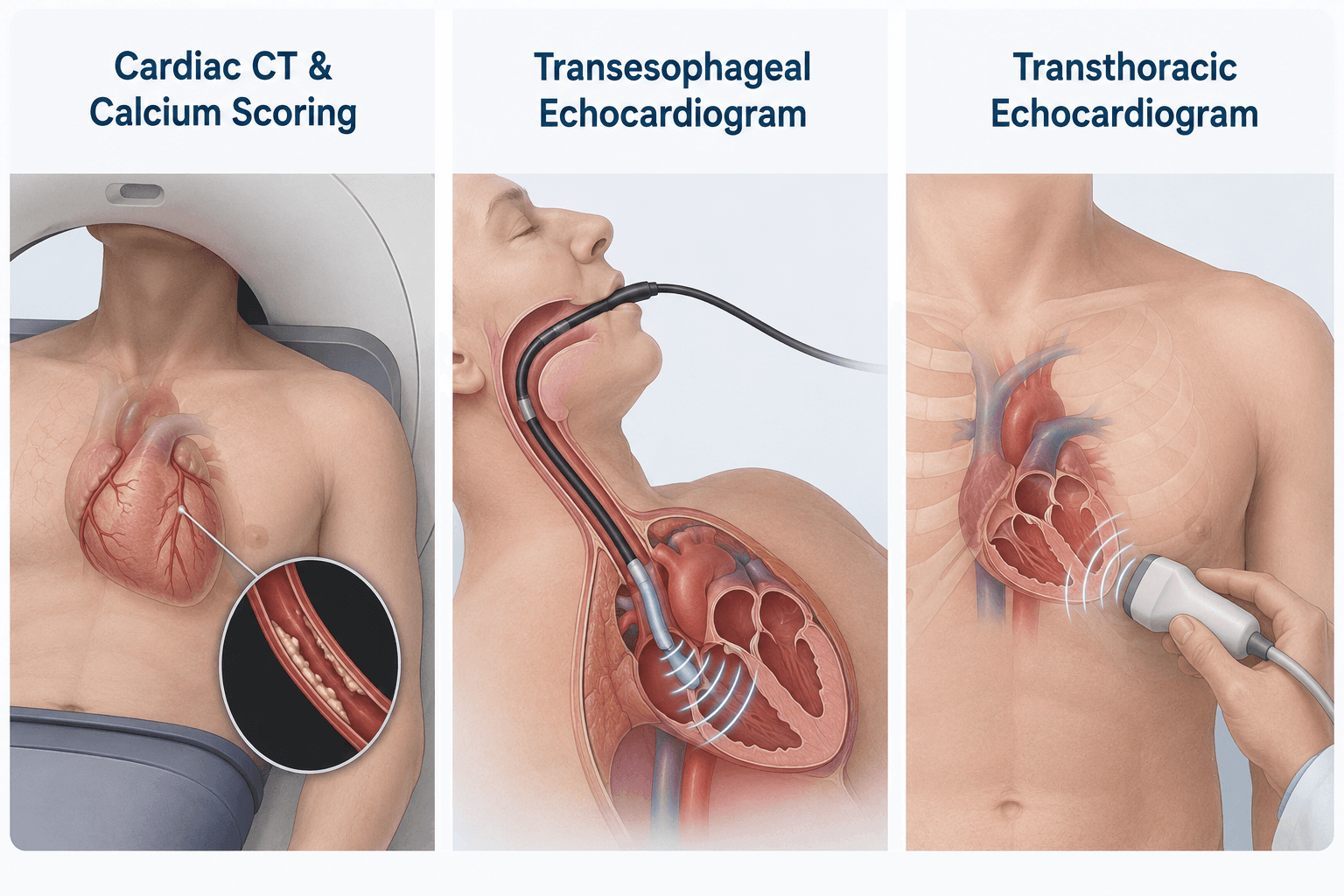
Finding the right answers starts with understanding your heart. We use gentle, non-invasive imaging tests to assess how your heart valves open and close, measure blood flow, and determine the best care plan for your health.
Transthoracic Echocardiogram (TTE)
Standard 2D/3D non-invasive ultrasound to evaluate valve anatomy, leaflet mobility, and chamber dimensions.
Transesophageal Echocardiogram (TEE)
High-resolution specialized ultrasound via an esophageal probe, providing clear, unobstructed views of complex valve structures.
Cardiac CT & Calcium Scoring
Detailed cross-sectional imaging evaluating aortic valve calcification and annular dimensions prior to TAVR or surgical intervention.
Treatment
Modern treatments include both surgery and less invasive catheter-based options for selected patients.
Transcatheter Aortic Valve Replacement (TAVR)
An expandable artificial valve is deployed inside the diseased aortic valve via a small leg artery catheter puncture.
MitraClip™ Transcatheter Edge-to-Edge Repair
A small clip is guided into the heart to hold mitral valve leaflets together and reduce severe mitral regurgitation.
Balloon Aortic / Mitral Valvuloplasty
Catheter balloon inflation used to stretch open stiff, narrow heart valve openings in select symptomatic patients.
Surgical Valve Repair or Replacement
Traditional or mini-thoracotomy surgical repair or replacement with mechanical or tissue prosthetic valves.
The main goal is to protect heart function and quality of life by monitoring carefully and treating at the right time—not too early or too late.