Thromboembolic
Disease & VTE
Thromboembolic Guide
Experiencing Sudden Breathlessness or Clot Symptoms?
Book EvaluationExperiencing Sudden Breathlessness or Clot Symptoms?
What is Thromboembolic Disease?
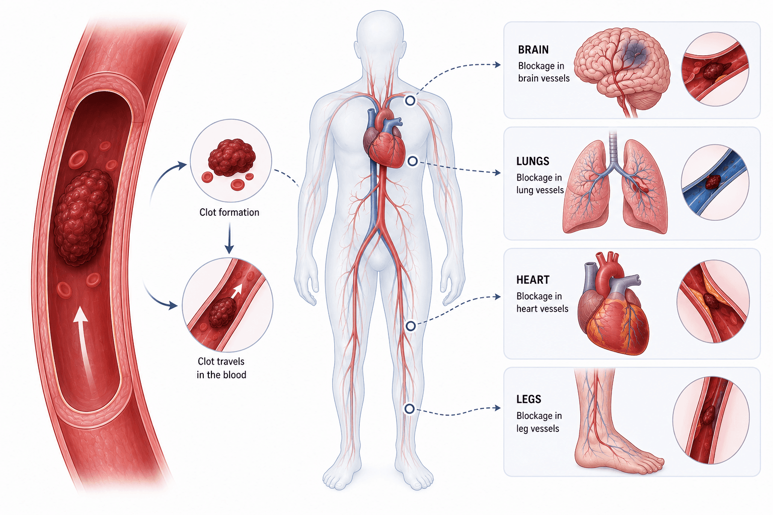
Thromboembolic disease occurs when blood clots (thrombi) form inside venous blood vessels. If a clot breaks loose from lower limb deep veins, it travels through the vena cava and right heart chambers into pulmonary lung arteries — causing a potentially catastrophic Pulmonary Embolism (PE).
What Patients Should Know
A clot is often a warning, not just an isolated event. When a DVT or PE occurs, it is important to understand why it happened—including recent surgery or immobilization, medications, cancer, inherited or acquired clotting disorders, or other risk factors.
Pulmonary embolism can be unpredictable. A relatively small clot may cause significant symptoms in some patients, while others may have few warning signs before a potentially serious event.
Symptoms of DVT & Pulmonary Embolism
Unilateral Swelling & Calf Warmth
Sudden, unexplained swelling in one leg accompanied by warmth and tenderness along the deep vein pathway — classic DVT presentation.
Sudden Shortness of Breath (Dyspnea)
Rapid onset of unexplained breathlessness occurring at rest or with minimal effort — hallmark PE symptom. Can be the only symptom.
Sharp Pleuritic Chest Pain
Stabbing chest pain that worsens significantly with deep inspiration or coughing, due to pulmonary infarction near the pleural surface.
Tachycardia & Rapid Pulse
Elevated heart rate over 100 beats per minute triggered by pulmonary vascular obstruction and right ventricular strain.
Hemoptysis (Coughing Blood)
Blood-tinged sputum resulting from pulmonary infarction — hemorrhagic necrosis of lung tissue distal to the occluded artery.
Syncope & Near-Fainting
Massive PE can cause sudden cardiovascular collapse as the right ventricle fails acutely from acute pressure overload.
Diagnostic
Diagnosing thromboembolic disease starts with understanding your symptoms, medical history, and risk factors. We use a clinical assessment and targeted tests, such as ultrasound and CT imaging, to determine whether a clot is present, where it is located, and how it is affecting blood flow.
CT Pulmonary Angiography (CT-PA)
Visualizes thrombi directly in pulmonary arteries with high resolution and accuracy to confirm or rule out a pulmonary embolism.
Lower Extremity Duplex Ultrasound
High-resolution vascular ultrasound evaluating deep leg veins to detect or rule out deep vein thrombosis (DVT).
D-Dimer Blood Assay
Measures clot-degradation protein fragments in the blood to help rule out active clot formation in appropriate patients.
Echocardiogram
Assesses right ventricular strain, heart chamber function, and pulmonary artery pressures affected by blood clots.
Treatment
Treatment focuses on stopping the clot from growing, preventing new clots, and reducing the risk of complications. Depending on the location and severity of the clot, treatment may include blood-thinning medications, clot-removal procedures, or clot-dissolving treatment in selected cases.
Direct Oral Anticoagulants (DOACs)
Apixaban, Rivaroxaban, Dabigatran. First-line therapy for most VTE. No routine INR monitoring. Proven to be as effective as warfarin with significantly lower bleeding rates.
Low Molecular Weight Heparin (LMWH)
Enoxaparin subcutaneous injections. Preferred for cancer-associated VTE (LMWH or DOAC) and during pregnancy (DOACs cross the placenta).
Catheter-Directed Thrombolysis (CDT)
Low-dose local tPA delivered directly into the pulmonary artery clot via catheter. For submassive PE with RV dysfunction — maximizes efficacy while minimizing bleeding risk.
IVC Filter Placement
Retrievable inferior vena cava filter placed when anticoagulation is absolutely contraindicated. Prevents clot migration from legs to lungs.
Preventing Blood Clots
Stay Mobile on Long Flights
Walk the aisle every 1–2 hours. Perform ankle pumping exercises while seated. Stay well hydrated.
Graduated Compression Stockings
15–30 mmHg stockings during travel or prolonged standing reduce DVT risk by 50%.
Prophylactic Anticoagulation
All high-risk surgical patients receive LMWH or fondaparinux injections pre- and post-operatively.
Early Mobilization After Surgery
Getting out of bed within 24 hours of surgery dramatically reduces the risk of DVT from venous stasis.